Plantar Fasciitis Review
Everything You Need to Know About Plantar Fasciitis
Guide to Recovery
What Is Plantar Fasciitis?
Plantar Fasciitis occurs when there is inflammation in the plantar fascia, particularly at the origin of the Flexor Digitorum Brevis Muscle (located at the medial process of the Calcaneal Tuberosity). It is associated with heel pain as well as pain along the inner aspect of the foot particularly during weight bearing movements.
What Causes Plantar Fasciitis?
Chronic overuse of the plantar fascia cause pain in the foot. Activities such as prolonged repetitive walking or running can cause micro-trauma to the involved tissues, leading to chronic inflammation and pain. This tends to occur when the rate of micro-trauma exceeds the rate of tissue recovery. During the gait cycle, tension increases in the plantar fascia to store potential energy during the Heel-Lift stage. This energy is then converted into kinetic energy at the Toe-Off stage to cause passive contraction of the plantar fascia. This mechanism creates foot acceleration to produce efficient locomotion.
The Windlass Effect occurs when the toes extend. This causes a stretch in the plantar fascia over the metatarsals (similar to how a pulley would work). This stretch causes a tension of the plantar fascia, and also shortens the longitudinal arches of the foot. This creates a spring mechanisms during Toe-Off.
Due to both the mechanism of the Windlass Effect and the location of the plantar fascia’s origin, pain at the heel can occur during both Heel-Lift and Toe-Off. With repetitive overuse of the plantar fascia, the constant irritation of the fibers can result in a degenerative process that may not involved inflammation at all (but still produce pain). In which case it is proposed that the name Plantar Fasciosis is a more appropriate diagnosis.
Who Is Most Affected By Plantar Fasciitis?
This condition is considered to be very common with a rough incidence being 15% of all foot injuries. This conditions tends to occur in younger runners, or adults aged 30-50 years old. There is a 2:1 ratio in occurrence in females over males.
Some risk factors include:
Ankle Dorsiflexion Hypomobility
Occupations requiring prolonged standing or walking (Warehouse workers, cashiers, servers)
Excessive Foot Pronation (Pes Planus)
Heel Spurs
Over training - Runners, Dancers, Track
Sudden increase in exercise intensity or duration - New Runners or Exercisers
Poor Foot Wear
Older Age (due to the natural decrease in the heel’s fat pad)
Sign and Symptoms of Plantar Fasciitis
Gradual increase in sharp pain in the heel
Pain is worse after rest (first few steps in the morning)
Feels better with light activity but worse with moderate to heavy activities
Pain is not constant and decreases with rest
Co-existing history of new shoes, increased running activities, or new job / work activities
How to Test for Plantar Fasciitis
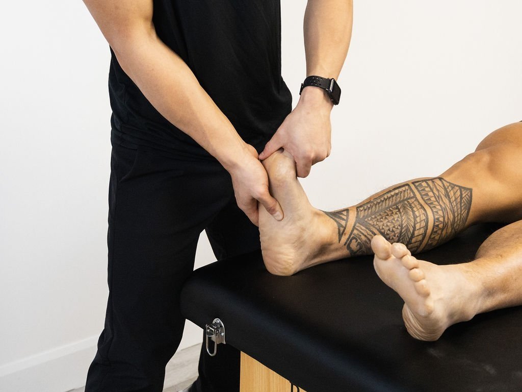
The Calcaneal Squeeze test is used to diagnose this condition. The diagnostician will place their thumb along the calcaneal insertion of the flexor digitorum brevis muscle and compress it to see if pain is reproduced.
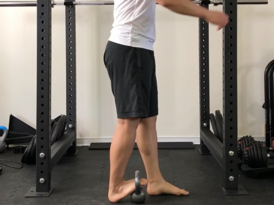
The Weight Bearing Windlass Test is also used to assess for Plantar Fasciitis. The examiner will get the patient to stand on their feet and extend the toes to stretch out the plantar fascia. A positive test is indicated when pain is reproduced at the calcaneus.
Do I Need an X-Ray?
An x-ray may be used to assess for any co-existing symptomatic calcaneal bone spurs. However it should be known that bone spurs are often asymptomatic (do not cause pain) and can be present in normal populations.
MRIs may be used to verify the location of any acute tears in the fascia, as well as identify local swelling. However MRIs are not required to make a clinical diagnosis
Diagnostic Ultrasound may be used to show hypertrophy of the fascia (increase in plantar fascia size). However it is not recorded to make a diagnosis.
Types of Treatments For PF
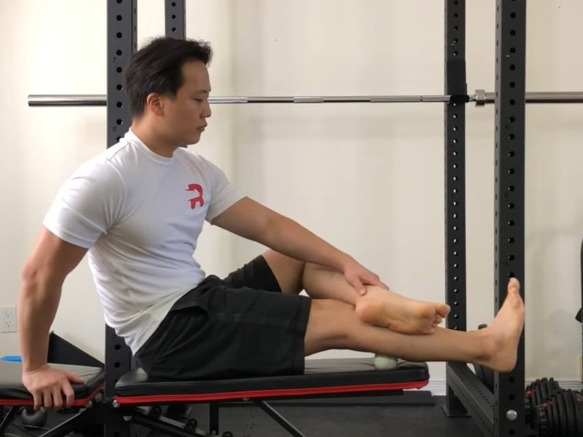
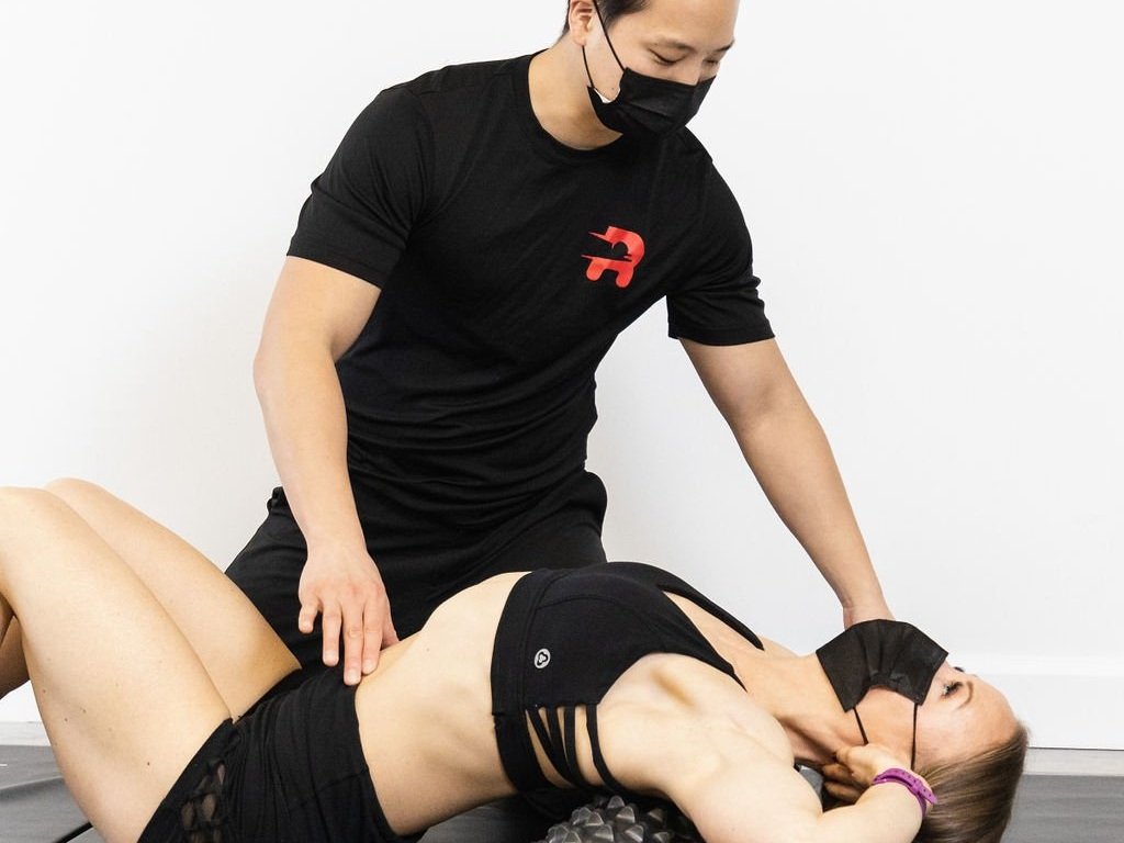
Conservative management is often indicated for this diagnosis. This may include stretches for the plantar fascia (non-weightbearing and weightbearing). Massage therapy can be applied through myofascial release or instrument assisted release techniques to both the plantar fascia and calf muscles (gastrocnemius and soleus). Joint mobilization or manipulation by a chiropractor or physiotherapist can be helpful for identified biomechanical compensations. This may include joints a the foot, ankle, knee, hip, or low back.
Heel pads may also be used if indicated for symptomatic heel spurs.
Modalities such as extracorporeal shock-wave therapy has been shown to be effective for plantar fasciitis as an adjunct therapy.
Acupuncture may also be applied in both the foot (near the calcaneus) and calf muscles.
Do I Need Surgery?
Surgery is very rarely used to manage this condition. This option is considered only after consistent conservative management has failed after approximately 1 year of treatments.
What Are Some Rehabilitation Exercises For PF?
Rehabilitation will be specific to what activities you can handle. A healthy combination of mobility exercises and strengthening exercises will be important for full recovery. For a full strengthening program consult your local sports physiotherapist or chiropractor in-person or virtually (online). Use the button below to book either appointment with a Rehab Hero therapist:
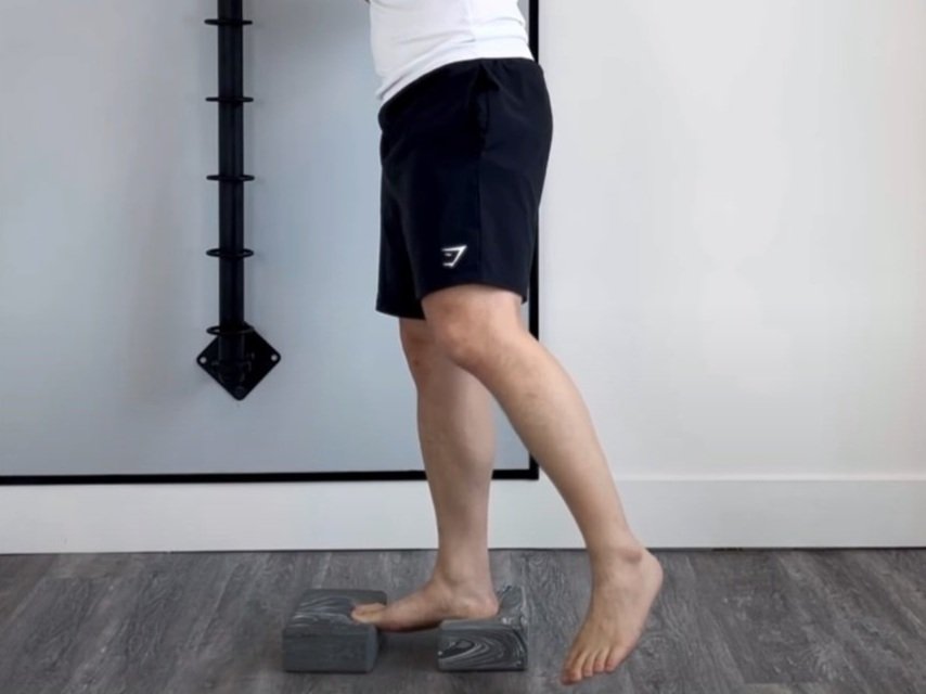
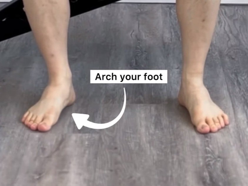
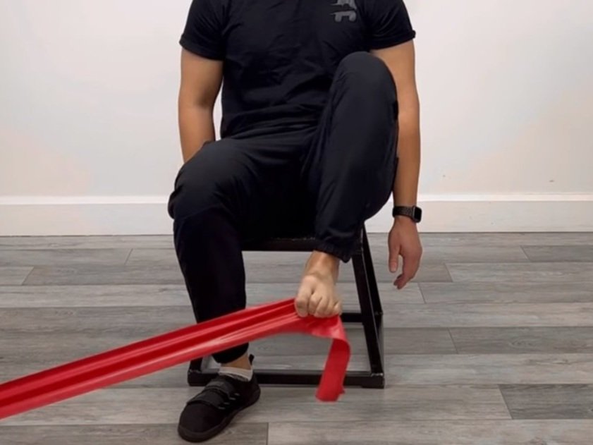
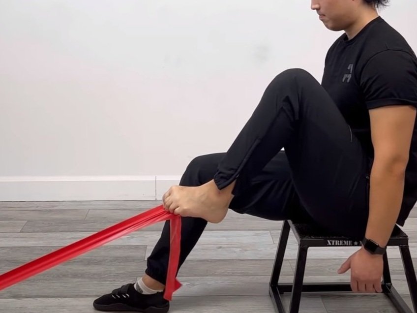
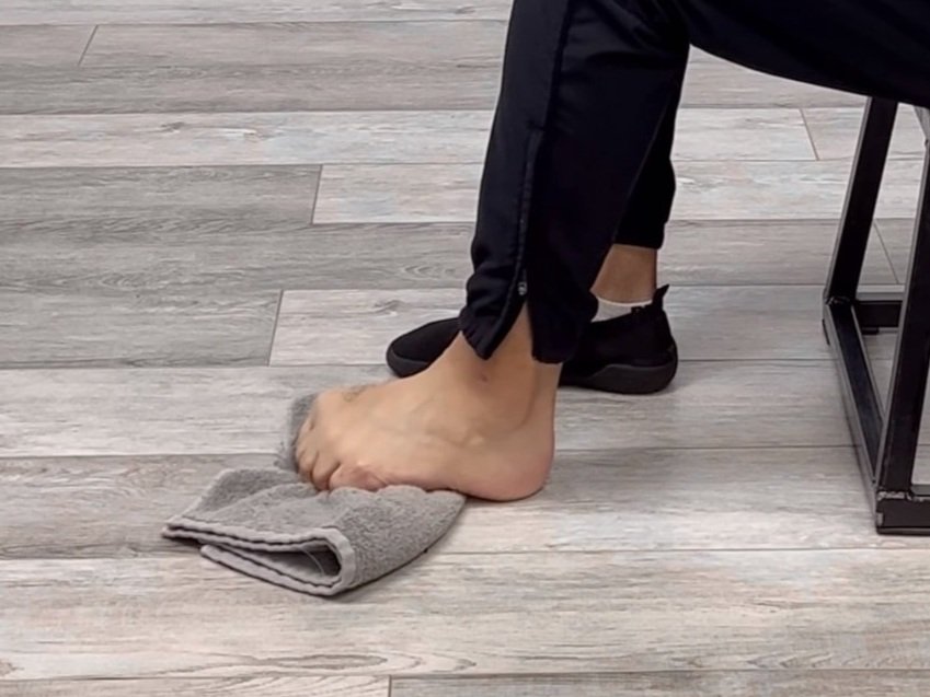
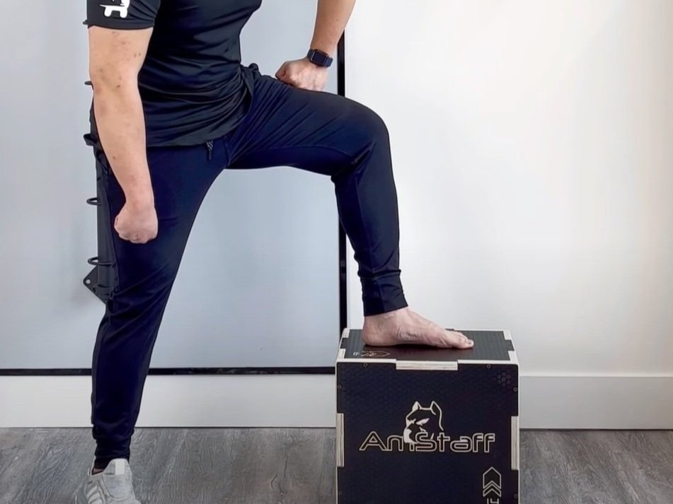
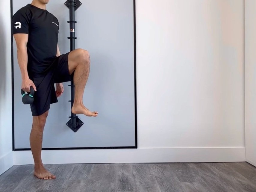
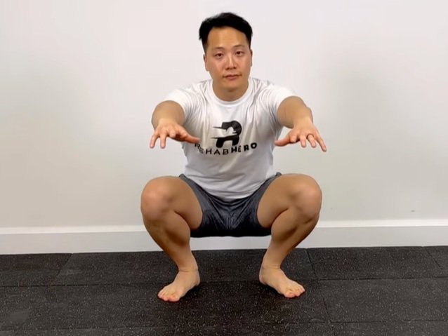
Some early care symptom management exercises are:
To watch a full video with exercise descriptions on how to start doing beginner exercises for plantar fasciitis click play below to learn from a Toronto chiropractor.
Other Pro Tips
With regards to managing plantar fasciitis, reducing aggravating activities will be the most important measure in creating a window of time where progressive exercises can occur. Other factors include:
Wearing correct foot wear
Walking and running biomechanics training
Ergonomic work correction

