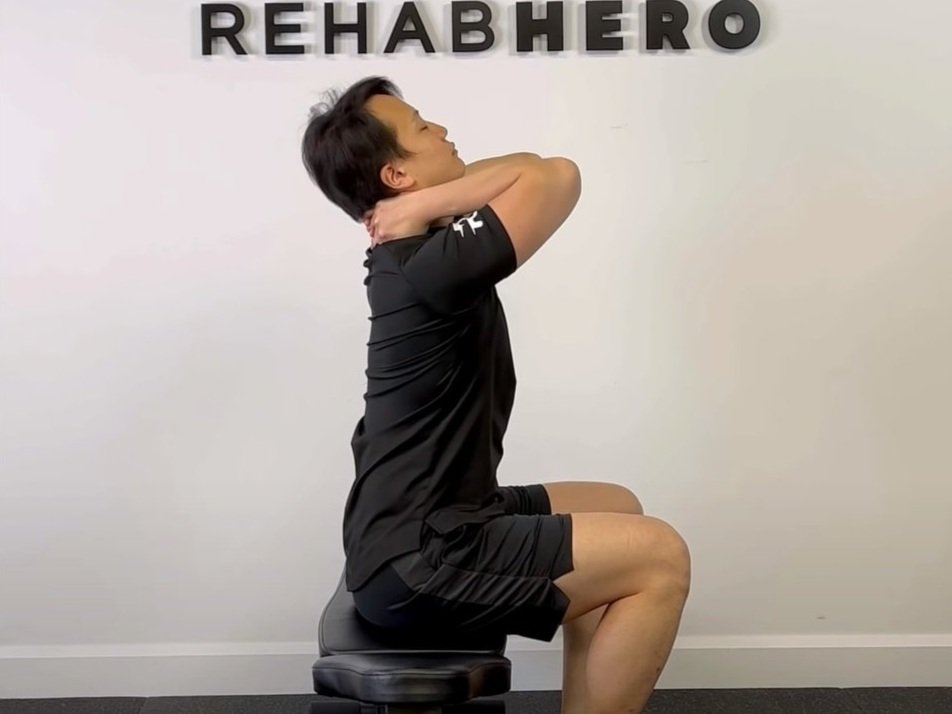
First Rib Dysfunction Review
First Rib Dysfunction and Neck Pain
Differential Diagnosis for Upper Trapezius Pain
The First Rib & Local Pain Generators
The first rib can become a pain generator for neck pain and is commonly found in individuals under the age of 50. Symptoms tend to be a result of superior translation of the first rib. Symptoms may also involve the shoulder and arm and persist as chronic neck or brachial pain. There are many sensitive structures that lie within the vicinity of the first rib, which include the sympathetic (stellate) ganglion, T1 ventral ramus, lower trunk of the brachial plexus, first intercostal nerve and vessels, and subclavian artery. Superior translation of the first rib can cause mechanical or inflammatory irritations to these local structures. Associated conditions also include thoracic outlet syndrome, and reflex sympathetic dystrophy.
The First Rib and The Scalene Muscles
Elevation of the first rib is known to occur due to the pull of the scalenes muscles, with superior translation of a rib being unique to the first rib. This condition may be cyclical in that there is a reactive response to contract during superior translation by the scalenes. Superior translation may occur with anterior or posterior translation depending on the involvement and pull of the scalenes muscles. Superior translation of the first rib may occur with elevation at the costovertebral and costotransverse joints, with some sources stating that superior translation occurs with superior elevation of the rib shaft and inferior translation of the rib head at the costovertebral joint. Sources also claim that the superior translation of the first rib is maintained due to a catching mechanism on the transverse process of T1, and therefore reduction will require an anterior and lateral translational force to dislodge the first rib. However, tension on the scalenes may resist reduction by preventing anterior translation of the first rib during reactive contraction.
First Rib Pathomechanics
Superior translation of the first rib is considered a structural dysfunction that may have resulted from hypertonicity of the scalenes, upper thoracic spine dysfunction, or lower cervical spine dysfunction. Cervical spine restrictions is hypothesized to cause irritation of the first rib through its innervation of the scalenes (nerve roots C3-C8), which may cause the scalenes to spasm and thus elevate the rib. Dysfunction of the first rib may also occur with forceful pushing or pulling with the arm while the elbow is locked in extension, this allows forces to transmit through the arm to the first rib. Treatment of these adjacent areas may help to relieve symptoms experienced.
This structural dysfunction may also cause a respiratory dysfunction such as restricting inhalation and exhalation. This can be examined by palpating the first ribs bilaterally and examining its movement during full respiration, restrictions may be found with earlier cessation of movement on the symptomatic side.
Clinical Presentation of First Rib Dysfunction
Clinical presentation includes pain palpated locally at the costovertebral, costotransverse or costosternal joints of the first rib, with or without referred pain to the head, neck and arm. Restricted motion during breathing may be palpated during either inspiration or expiration. Rib springing may feel asymmetrical compared to the asymptomatic side. Palpable displacement, tenderness, or swelling may be revealed at the head, at the sternal end, or at the superior anterior surface of the supraclavicular fossa. Cervical contralateral lateral flexion and ipsilateral rotation will be positive for pain or limitation. Associated findings include restrictions in the cervical or thoracic spine. Cervical extension will reproduce symptoms and may be described as a pulling sensation. Shoulder movement may also be painful at end ranges as well.
Additional associated findings include reproduction of symptoms during unilateral posteroanterior pressure of the first rib or at it’s costotransverse joint. Pain may also be reproduced with caudal pressure on the anterior or lateral aspect of the first rib. It is not uncommon to have upper trapezius spasm that is tender on palpation as well. Paresthesia and dysthesia of C8 and T1 nerve root distributions have also been reported, with or without signs of reflexive sympathetic dystrophy (sweating, discolouration, swelling, clumsiness, weakness at the hand and forearm).
Who Should I See to Get Diagnosed?
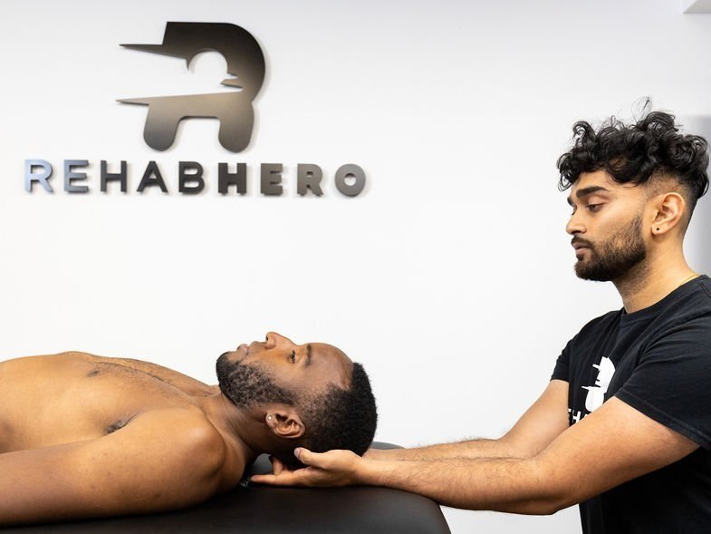
You can get diagnosed by a physiotherapist or chiropractor and in Ontario no referral from a medical doctor is required to book an appointment. During your chiropractic or physiotherapy appointment, your clinician will test your overall movement in the neck, shoulder, and upper back regions. This information will be used in combination with orthopaedic and neurological tests to rule out other conditions that present similarly. Your health care provider will also take a look at muscle tone and joint mobility with palpation and compare side to side to look for any relevant asymmetries.
First Rib Treatment Options
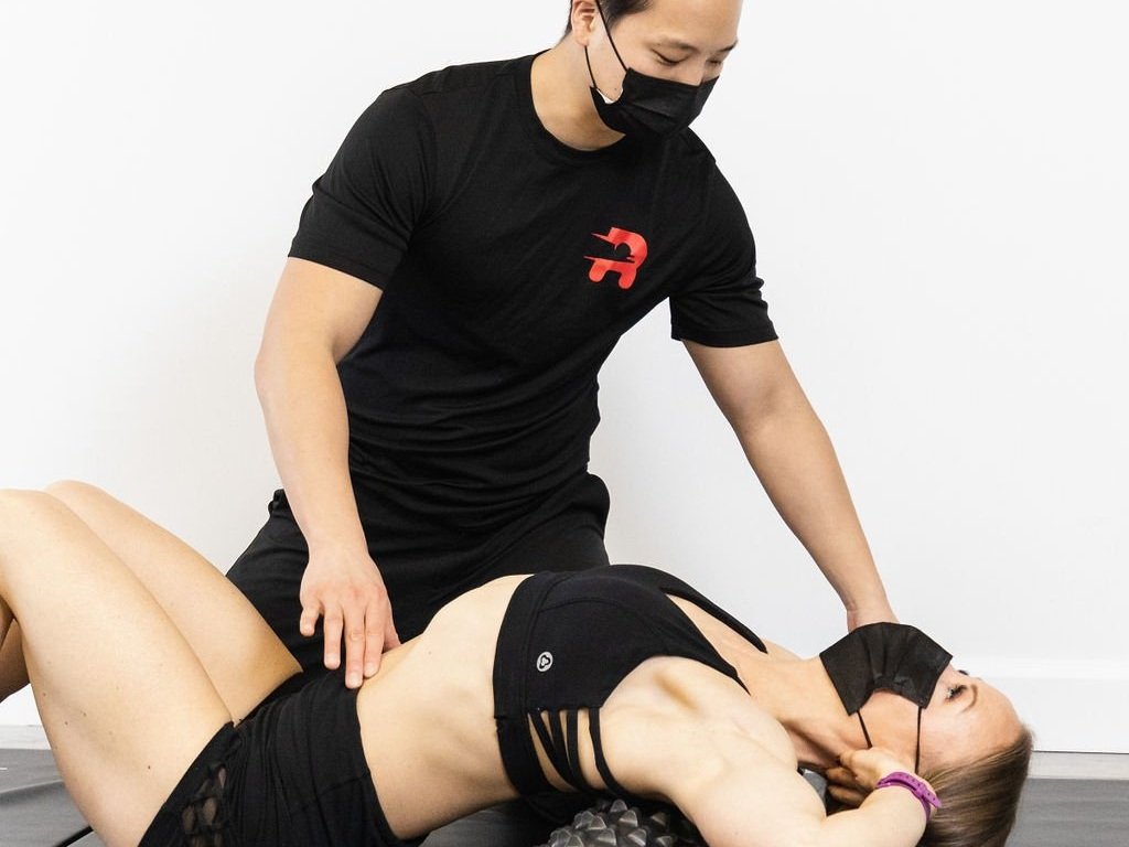
Based on your clinical findings, your physiotherapist or chiropractor will use a combination of manual therapy and rehabilitative exercises. Manual therapy may include joint mobilizations of the costotransverse, costovertebral, cervical facet, or thoracic facet joints. It may also include myofascial release of the scalenes, upper trapezius, levator scapulae, splenius capitus, splenius cervicis, or sternocleidomastoid muscles to name a few. Rehabilitative exercises will generally be focused on breathing mechanics, thoracic mobility exercises, cervical stability exercises, as well as dissociative exercises between the shoulder, neck and back. Adjunctive therapies include cupping, acupuncture, cold laser and kinesiotaping.
Exercises For First Rib Dysfunction
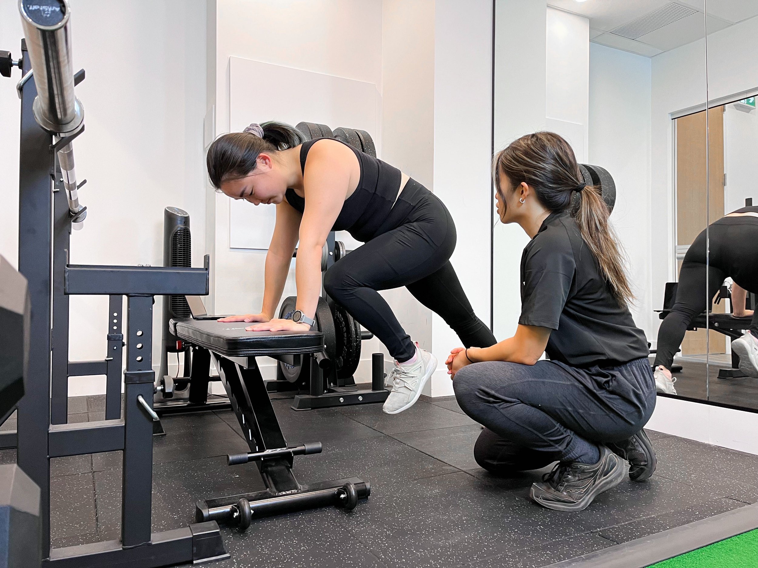
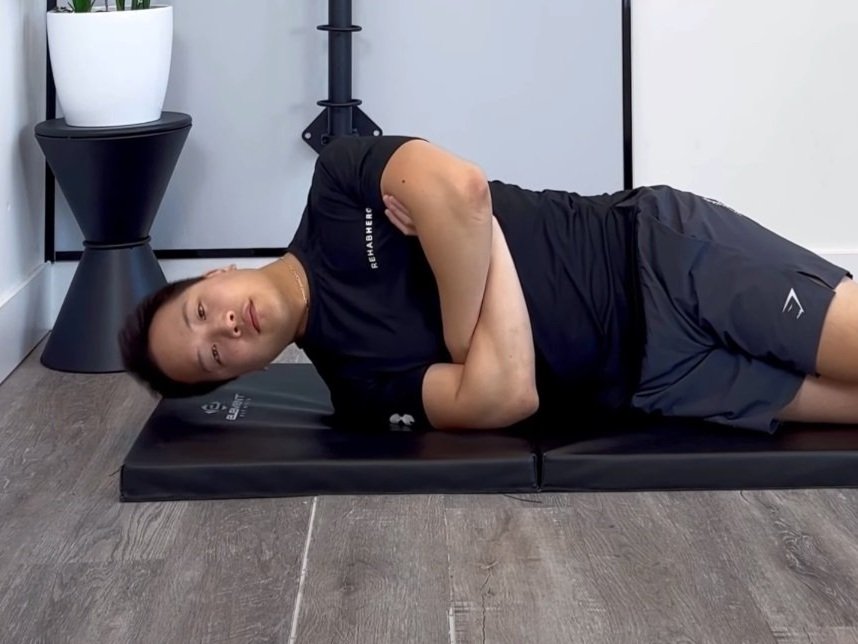
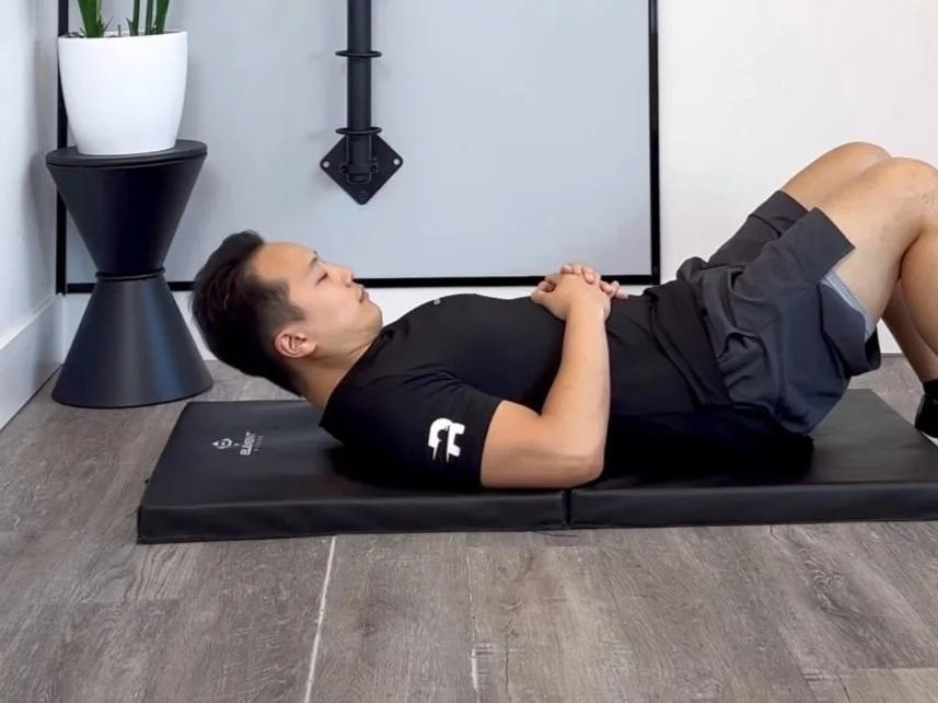
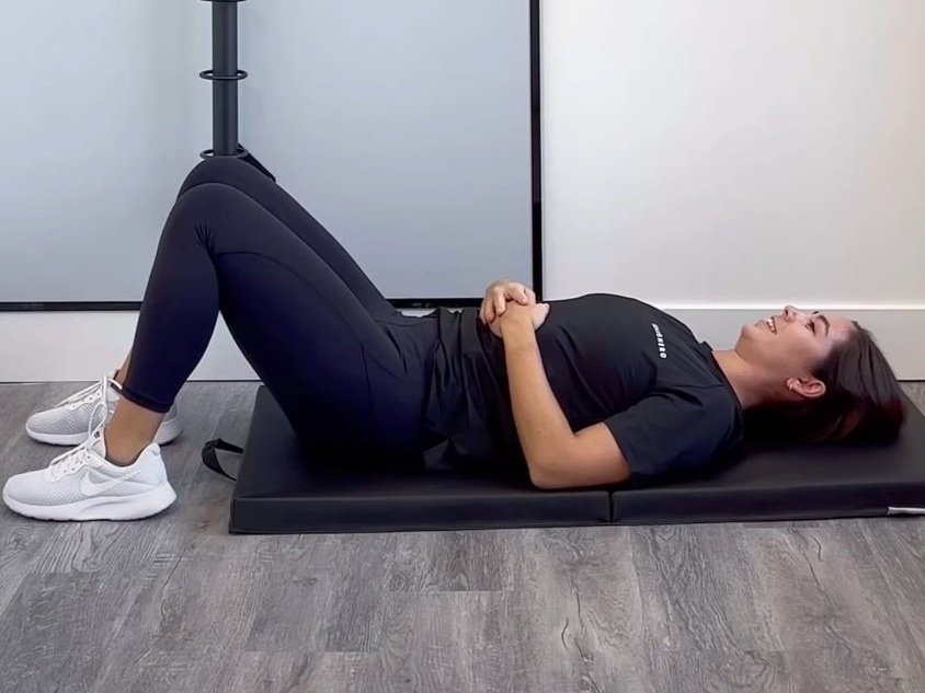
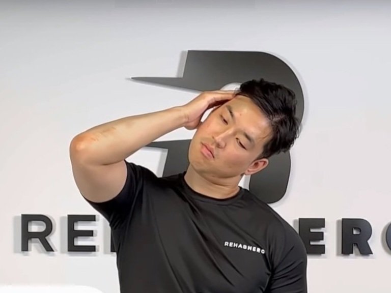
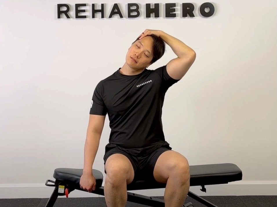
Exercises are typically prescribed based on clinical findings and will be specific to your movement assessment. Although the diagnosis may be the same, individual circumstances and movement capacity will defer from person to person. Generally exercises that target thoracic mobility and cervical stability are used to improve patient outcomes. Exercises may include:
**DISCLAIMER: Please note that it is recommended to consult a health care provider about your diagnosis prior to trying out any new exercises. Circumstances vary and these exercises may not be right for you, to prevent injury seek out a professional for help. Perform these exercises are at your own risk.**
Check out this full length video on how to recover from an elevated first rib:
Written By:
Dr. David Song, Chiropractor, Rehab Coach
References
1. Kamkar A, Cardi-Laurent C, Whitney S. Conservative management of superior subluxation of the first rib. Journal Of Sport Rehabilitation [serial on the Internet]. (1992, Nov), [cited November 7, 2017]; 1(4): 300-316. Available from: SPORTDiscus with Full Text.
2. Brismée J, Phelps V, Sizer P. Differential diagnosis and treatment of chronic neck and upper trapezius pain and upper extremity paresthesia: a case study involving the management of an elevated first rib and uncovertebral joint dysfunction. Journal Of Manual & Manipulative Therapy (Journal Of Manual & Manipulative Therapy) [serial on the Internet]. (2005, June), [cited November 7, 2017]; 13(2): 79-90. Available from: CINAHL Plus with Full Text.


